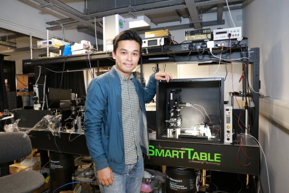
HKU biomedical engineers achieve significant breakthrough in neuroimaging with novel high-speed microscope to capture brain neuroactivities
Our brain contains tens of billions of nerve cells (neurons) which constantly communicate with each other by sending chemical and electrical flashes, each lasting a short one millisecond (0.001 sec). In every millisecond, these billions of swift-flying flashes altogether traveling in a giant star-map in the brain that lights up a tortuous glittering pattern. They are the origins of all body functions and behaviours such as emotions, perceptions, thoughts, actions, and memories; and also brain diseases e.g. alzheimer’s and parkinson’s diseases, in case of abnormalities.
One grand challenge for neuroscience in the 21st century is to capture these complex flickering patterns of neural activities, which is the key to an integrated understanding of the large-scale brain-wide interactions. To capture these swift-flying signals live has been a challenge to neuroscientists and biomedical engineers. It would take a high-speed microscope into the brain, which has not been possible so far.
A research team led by Dr Kevin Tsia, Associate Professor of the Department of Electrical and Electronic Engineering and Programme Director of Bachelor of Engineering in Biomedical Engineering of the University of Hong Kong (HKU); and Professor Ji Na, from the Department of Molecular & Cell Biology, University of California, Berkeley (UC Berkeley) offers a novel solution with their super high-speed microscope – two-photon fluorescence microscope, which has successfully recorded the millisecond electrical signals in the neurons of an alert mouse.
For details, please click here.


